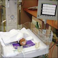
Till now the aEEG technique has been tested and validated in a variety of situations at different age groups, from adults anesthetized and undergoing cardioplegia during open-heart surgery (4) to preterm infants in an intensive care environment (5). The main usage of aEEG nowadays (as is in our unit (6)) is monitoring full term newborns after birth asphyxia and/or suspected of seizures. The signal output from this group of patients underwent extensive investigations (6-10) and brought the technique from the status of an investigational tool to one of an accepted monitoring technique in various neonatal intensive care units. Monitoring infants with the CFM after severe birth asphyxia is a quite accurate mean for prognosis prediction (7-10, 14, 16) and it can influence various treatment modalities. Another well established use of the CFM is monitoring for seizure activity (11, 12, 15), both clinical and silent with an added advantage of monitoring the treatment effect (13). In the various works including ours (17) a good correlation was noted between aEEG and conventional EEG signal, pointing out that the aEEG can reliably detect seizures (though short, focal and low amplitude seizures might be missed). Thus, the main advantages of this technique are its simplicity and ease of both application and interpreting on one hand and on the other the possibility of continuous long term monitoring with real time assessment of clinical events.
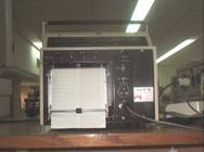
Connection instructions in hebrew for the analog Lectromed prototype
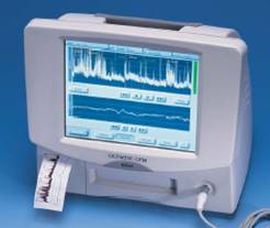
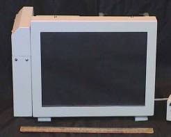
Connection instructions in hebrew for the digital Olympic CFM 6000 prototype
Connection instructions in english for the digital Olympic CFM 6000 prototype
Brain monitors. This site gives some information about the history of the CFM and newer versions called CFAM.
CFM 6000 home page. This is the official Olympic Medicals web site of a new digital CFM (CFM 6000).
BRM2 home page. This is the official BrainZ Instruments web site of a new digital CFM (BRM2).
Bibliography:
1. Prior PF, Maynard DE, Sheaff PC et al, Monitoring cerebral function: clinical experience with a new device for continuous recording of electrical activity of brain. Br Med J, 1971; 2: 736-738. back
2. Maynard DE, Prior PF and Scott DF, Device for continuous monitoring of cerebral activity in resuscitated patients. Br Med J, 1969; 4: 545-546. back
3. Hellstrom-Westas L, Cerebral function monitoring in neonatal intensive care. Thesis, department of pediatrics, University of Lund, 1990. back
4. Silvay G, Mindich BP, Owitz S, Koffski RM, Litwak RS, Evaluation of a new cerebral function monitor during open-heart surgery. Mt Sinai J Med, 1983; 50: 44-48. back
5. Hellstrom-Westas L, Rosen I and Swenningsen NW, Cerebral function monitoring during the first week of life in extremely small low birthwheight (ESLBW) infants. Neuropediatrics, 1991; 22: 27-32. back
6. Shany E, Shorer Z and Karplus M, Cerebral function monitoring after severe perinatal asphyxia, Harefuah; 1998; 135; 440-445. back
7. Thornberg E and Ekstrom-Jodal B, Cerebral function monitoring: a method of predicting outcome in term neonates after severe perinatal asphyxia. Acta Paediatr 1994; 83: 596-601. back
8. Hellstrom-Westas L, Rosen I and Swenningsen NW, Predictive value of early continuous amplitude integrated EEG recordings on outcome after severe birth asphyxia in full term infants. Arch Dis Child 1995; 72: F34-F38. back
9. Bjerre I, Hellstrom-Westas L, Rosen I and Swenningsen N, Monitoring of cerebral function after severe asphyxia in infancy. Arch Dis Child 1983; 58: 997-1002. back
10. Toet MC, Hellstrom-Westas L, Groenendaal f, eken P and de Vries LS, Amplitude integrated EEG 3 and 6 hours after birth in full term neonates with hypoxic ischemic encephalopathy. Arch Dis Child Fetal Neonatal Ed 1999; 81: F19-F23. back
11. Hellstrom-Westas L, Rosen I and Swenningsen NW, Silent seizures in sick infants in early life, diagnosis by continuous cerebral function monitoring. Acta Paediatr Scand 1985; 74:741-748. back
12. Altfullah I, Asaikar S and Torres F, Status epilepticus: clinical experience with two special devices for continuous cerebral monitoring. Acta Neurol Scand 1991; 84: 374-381. back
13. Hellstrom-Westas L, Westgren U, Rosen I and Swenningsen NW, Lidocaine treatment of severe seizures in newborn infants, I. Clinical effects and cerebral electrical activity monitoring. Acta Paediatr Scand 1988; 77: 79-84. back
14. Niran al Naqeeb, MRCP, A. David Edwards, FRCP, Frances M. Cowan, PhD, and Denis Azzopardi, FRCP, Assessment of Neonatal Encephalopathy by Amplitude-integrated Electroencephalography. Pediatrics 1999; 103(6): 1263-1271. back
15. Shany E, Khvatskin S, Golan A, Karplus M, Amplitude-integrated electroencephalography: a tool for monitoring silent seizures in neonates. Pediatr Neurol. 2006 Mar;34(3):194-9. back
16.Shany E, Goldstein E, Khvatskin S, Friger MD, Heiman N, Goldstein M, Karplus M, Galil A. Predictive Value of Amplitude-Integrated Electroencephalography Pattern and Voltage in Asphyxiated Term Infants. Pediatr Neurol 2006; 35:335-342.back
17.Frenkel N, Friger M, Meledin I, Berger I, Marks K, Bassan H, Shany E. Neonatal seizure recognition--comparative study of continuous-amplitude integrated EEG versus short conventional EEG recordings. Clin Neurophysiol. 2011 Jun;122(6):1091-7. back
Connection instructions in hebrew
This Page was prepared by Dr. Shany Eilon
back to Eilon home page
| |||||
